Technologies

Murder the Result of Various Injuries, 1898
Suicide by Cutting the Throat, 1898
Encircling Gunshot-wound in Brain, 1898
Carbonic-oxide [carbon monoxide] Poisoning with agonal injuries due to a fall, 1898
Suicide through Stabbing, 1898
Bloodstain, blisters, bullet holes, 1864
Head and hand of a drownee, 1864
Decomposed stomach, 1864
Rope marks and upper thigh, 1864
The color of the lungs of dead newborn children: stillborn, newborn who have taken a breath, newborn whose lungs have been artificially inflated, 1864
Medical professor Johann Ludwig Casper's richly colored lithographic plates illustrate specific postmortem examinations, some of them experiments on cadavers.
Johann Ludwig Casper, M.D., Atlas zum Handbuch der gerichtlichen Medicin [Atlas for the Manual of Legal Medicine], Berlin; Artist: Hugo Troschel; Lithographer: Winckelmann & Sons
National Library of Medicine
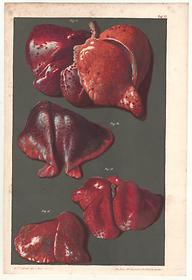
Image 10 of 10

The 19th-century revolution in forensic imaging

In the 19th century, forensic pathologists began using pictures and words to show how various conditions appear in the cadaver, and to teach students and colleagues new methods of analysis. Line drawings, half-tone photography, and chromolithography, which could render coloration, texture, and subtle shading, became increasingly common as improvements in print technology made detailed illustrations cheaper to produce.