Medical Views
Medicine and law overlap in a death investigation calling for an autopsy to determine the manner and cause of death. Listen to two chief medical examiners talk about their work and careers. Explore the medico-legal autopsy procedures, and find out what virtual autopsy is all about!
Medical examiners
Pathologists—medical doctors who specialize in anatomical or functional abnormalities of a human body—with a special forensic training work as medical examiners and conduct medico-legal autopsies. Meet Drs. Marcela Fierro and David Fowler, who speak about their career and experiences as the chief medical examiners of Virginia and Maryland, respectively.
 Becoming a chief medical examiner
View the video
Read the transcript
 Critical skills for medical examiners
View the video
Read the transcript
 Working with families
Autopsy
Explore the purposes of a medico-legal autopsy and learn about select autopsy pathologies from the two chief medical examiners' perspectives.
Autopsy overview
Why and how is an autopsy performed? Find out from the experts the general autopsy procedure and its purpose.
 General autopsy procedure
View the video
Read the transcript
 Purpose & criteria for a medico-legal autopsy
View the video
Read the transcript
Petechial hemorrhage
Take a close look at these eyes—look at your own to help you compare and note differences. Do you see the tiny red dots on the eye's upper white part and the inside of the eyelid? This condition is called petechial (tiny dots) hemorrhage (bleeding). What does this tell a medical examiner? When, during an autopsy, does a medical examiner find such a condition?
Petechial hemorrhage
 Petechial hemorrhage
View the video
Read the transcript
 Autopsy: external examination
View the video
Read the transcript
Polycystic kidney
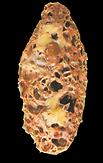
Here are the cross sections of a healthy kidney and one with adult polycystic kidney disease. Take a closer look at each and note their differences. What is polycystic kidney disease and how does it happen? How and when does a medical examiner examine kidneys during an autopsy?
Normal kidney cross section
Polycystic kidney disease
 Adult polycystic kidney disease
View the video
Read the transcript
 Autopsy: internal in situ examination
View the video
Read the transcript
Perforated heart
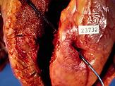
This heart has a perforated wound caused by a bullet. What does such a wound tell a medical examiner? How and when does she or he examine the heart?
Perforated heart
 Gunshot wound in the heart
View the video
Read the transcript
 Autopsy: organ examination
View the video
Read the transcript
Cerebral meningitis
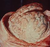
This brain shows an abnormal condition—yellow-tan clouding of the meninges, the three layers of membranes covering the brain—caused by a viral or bacterial infection. What does a medical examiner do with this finding? How does the infection affect people conducting the autopsy?
Brain with acute meningitis
 Fatal cerebral meningitis
View the video
Read the transcript
 Autopsy: brain examination & occupational hazard
Virtopsy
Find out how high-tech imaging technologies are applied to a medico-legal autopsy in searching for the cause and manner of death.
 What is virtopsy and what are its benefits?
View the video
Read the transcript
A research case
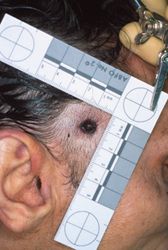
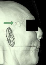
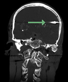
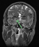
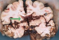
Drs. Richard Dirnhofer and Michael J. Thali and their team of specialists at the University of Bern's Institute of Forensic Medicine in Switzerland use multi-slice computed tomography (MSCT) and magnetic resonance imaging (MRI) in comparison with a forensic autopsy to determine how well MSCT and MRI can document and analyze gunshot wounds. Compare the photographic, MRI, and CT images from a case and explore how imaging technology applications may assist and supplement traditional autopsy.
Photograph of an entrance wound
3D CT surface reconstruction of the face showing a small entrance wound of the right temple (arrow)
Coronal CT reformation showing the projectile on the left (arrow)
MRI cross section (corresponding to the coronal CT reformation image) showing brain tissue details and the bullet track
Photograph of the brain section showing the bullet track and projectile
Medical Views is based on the "Autopsy Revealed" multimedia interactive station at the Visible Proofs: Forensic Views of the Body exhibition on display at the National Library of Medicine, Bethesda, Maryland from February 16, 2006–February 16, 2008. Visitor and guided-tour information is available online at VISIT. If you have questions about visiting Visible Proofs, contact the NLM Support Center.


