
Digital Gallery
Explore a curated selection of anatomical texts and illustrations from 1500s to the early 1900s, from the historical collections of the National Library of Medicine. Discover how anatomists' knowledge and perceptions influenced their depictions of the interior body.
31 Images
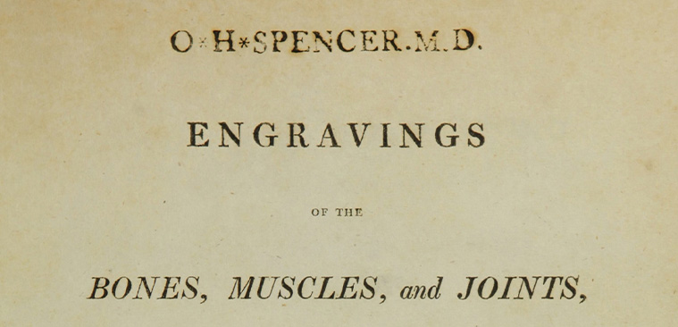
Engravings of the bones, muscles, and joints, illustrating the first volume of the Anatomy of the human body, John Bell, 1804
close next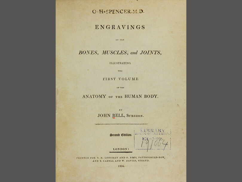
Scottish anatomist John Bell (1763–1820) drew, engraved, and etched all of his plates in the “Book of Bones,” the “Book of Muscles,” and the “Book of Joints.” His work diverged from other anatomy books of the period—which featured elaborate artistic and abstract representations of the body—and instead focused on simple, stand-alone anatomical renderings. He believed that traditional books distracted students’ ability to understand human anatomy. His illustrations depicted the tools and apparatuses used in dissection as well as the process of bodily decomposition, also essential knowledge for students.
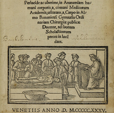
Anatomia Carpi. Isagoge breves perlucide ac uberime, in Anatomiam humani corporis, a communi medicorum academia usitatam, a Carpo in almo Bononiensi Gymnasio ordinariam chirurgiae publicae docente, ad suorum scholasticorum preces in lucemdate, Jacopo Berengario da Carpi, 1523/1535
close next previous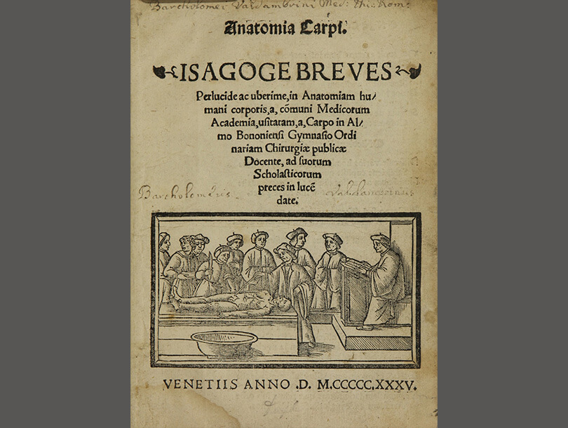
Italian surgeon and physician Jacopo Berengario da Carpi (ca. 1460–ca. 1530) was among the first to publish anatomy texts with illustrations based on his own dissection of hundreds of bodies. After the invention of the printing press in the 1450s, Berengario created multiple copies of anatomical woodcut engravings. In 1522, after studying the work of Italian surgeon and anatomist Mondino de Luzzi (ca. 1270–1326), he published Isagogae breves, a compilation of Mondino’s work that included Berengario’s woodcuts. This 1523 edition has four additional illustrations of the heart and the brain. Although the illustrations in this text are simple and do not contain significant anatomical details, Berengario’s work is considered the beginning of a new age in anatomical studies.
A compleat treatise of the muscles, as they appear in humane body, and arise in dissection; with diverse anatomical observations not yet discover’d ..., , John Browne , 1681
close next previous
English anatomist John Browne (1642–1702) became known for copying, altering, and presenting the illustrations of other engravers and anatomists as his own work. Although the legal concept of copyright did not yet exist, anatomists took notice. English surgeon and anatomist William Cowper, also accused of copying others, nevertheless openly criticized Browne for perpetuating errors in anatomy by copying outdated renderings. This text was also closely based on Myskotomia (1684) written by English anatomist and surgeon William Molins (1617–1691). Despite altering the poses, backgrounds, and clothing, many of the forty copper plate illustrations are nearly exact copies of Giulio Cesare Casserio’s (ca. 1552–1616)* illustrations in Tabulae Anatomicae (1627).
Myographia nova; sive, Musculorum omnium (in corpore humano hactenus repertorum) ... descriptio, in sex praelectiones distributa ..., John Browne, 1687
close next previous
Despite being accused of plagiarism after writing A compleat treatise of the muscles (1681), English anatomist John Browne’s (1642–1702) Myographia nova was yet another compilation of others’ work and also a successful publishing endeavor—10 editions were eventually printed. This particular edition contains a new syllabus musculorum by the anatomist Charles Scarborough (1615–1694) and additional copper plate illustrations. Publishing new editions of books in different languages with slightly modified content and new titles was a smart business decision for many of these scholars. By slightly altering new editions, Browne and other anatomists could keep selling books without producing much new work.
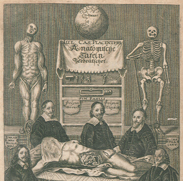
Anatomische Tafeln, Giulio Cesare Casserio Simon Paulli, 1656
close next previous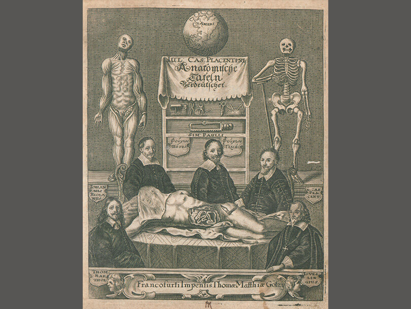
Anatomische Tafeln contains the German translation by Simon Paulli (1603–1680) of two well-known anatomical texts by the Italian anatomist Giulio Cesare Casserio (ca. 1552–1616) and his Belgian student, Adriaan van de Spiegel (1578–1625). The first is a reproduction of Tabulae anatomicae LXXIIX (1632) and the second is De formato foetu liber singularis (1626). Pauli also included appendix of Latin writings as the volume’s third part.
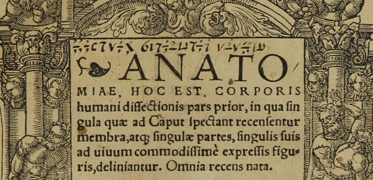
Corporis humani dissectionis pars prior, Johann Dryander, 1537
close next previous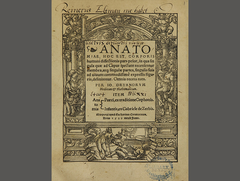
Anatomiae represents a transition to more modern anatomic descriptions and illustrations. Although influenced by the revered work of Italian anatomists Mondino de Luzzi (ca. 1270–1326) and Jacopo Berengario (ca. 1460–ca. 1530), German anatomy professor Johann Dryander (1500–1560) departed from traditional anatomical representations. Dryander was one of the first anatomists to base his illustrations on his own dissections, while most anatomists blurred the line between anatomy and art by presenting human corpses in classical art poses in their illustrations. This book contains 20 copperplate illustrations: 16 of the head and brain and 4 of the chest and lungs.
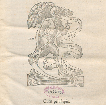
De dissectione partium corporis humani libri tres, Charles Estienne, 1545
close next previous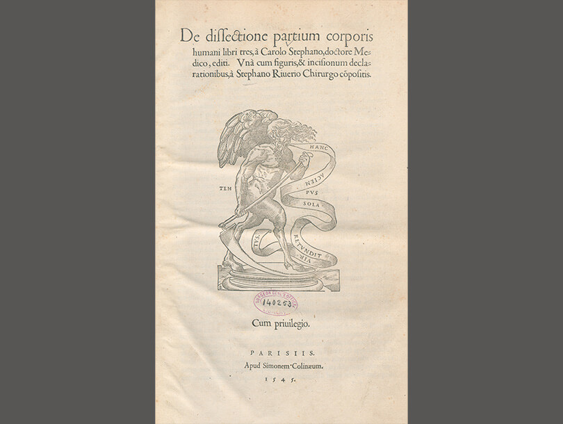
In a 1530s scholarly collaboration, French physicians Charles Estienne (1504–ca. 1564) worked with Etienne de La Rivière (d. 1569) on dissections and illustrations while artist Jean Jollat (fl. 1530–1545) produced the woodcuts. Although ready for publication in 1539, a dispute between the two delayed printing of the book until 1545. Meanwhile, in 1543, the Flemish anatomist Andreas Vesalius (1514–1564) published, De humani corporis fabrica libri septem.
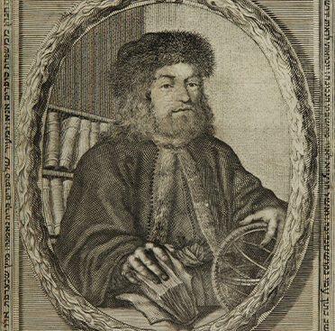
Ḥeleḳ sheni mi-Sefer Maʻaśeh Ṭoviyah, Ḥeleḳ shlishi mi-Sefer Maʻaśeh Ṭoviyah, and Sefer ha-ʻOlamot, Ṭoviyah Kats, ca. 1708
close next previous
Maʻaśeh Ṭoviyah, a Hebrew encyclopedia, is one of the first Hebrew books to have scientific and medical illustrations. Its frontispiece shows a portrait of the author, Ṭoviyah Kats (ca. 1652–1729), surrounded by books and wearing a doctor’s ring, a fur lined cape, and a hat. He holds a book in one hand, and an astroglobe in the other—symbolizing his quest to learn the natural order of the universe. The encyclopedia has eight sections—theology, astronomy, medicine, hygiene, illnesses from syphilis, botany, the universe, and the four elements—but most of it is devoted to medicine. One notable engraving quaintly compares the interior of a human body with that of a four-story house.
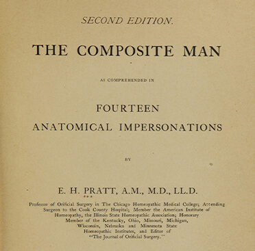
The composite man as comprehended in fourteen anatomical impersonations, Edwin Hartley Pratt, 1901
close next previous
The Composite Man first appeared as serial chapters in the Journal of Orifical Surgery between April 1899 and January 1901. Soon after, Edwin Hartley Pratt (1849–1930) compiled them into a book to popularize anatomy by using accurate, accessible, and humorous writing. Each chapter introduces a different “man” who explains his anatomical function to the reader. Some of these include: Bony Man, Muscular Man, Arterial Man, Venous Man, Lymphatic Man, Skin Man, Connective Tissue Man, Cerebro-Spinal Man, and Sub-Conscious Man. A simple anatomical color illustration of the featured “man” accompanies each chapter. He succeeded in his effort to popularize anatomy: the publisher printed 19 editions between 1901 and 1921.
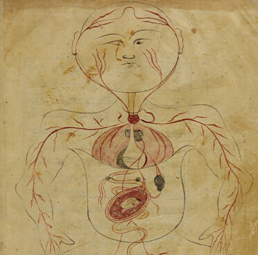
Tashrīḥ al-badan, Mansūr ibn Muhammad ibn Ahmad ibn Yūsuf ibn Faqīh Ilyās (author); Ardistānī, Hasan ibn Ahmad kātib-i muqīm-i Isfahān (scribe), 1488
close next previous
This rare Persian manuscript was one of the first in the world to contain detailed anatomical illustrations. The title translates to “Description of the Body,” and the text summarizes many of the observations made by the Ancient Greek physician, surgeon, and philosopher Claudius Galen (129–ca. 216). Six illustrations detail the spinal cord, circulatory and skeletal systems, skin, digestion, and human gestation. The final picture shows a female body carrying a fetus, but the woman’s body looks no different than all the other illustrations. Medieval Muslim physicians added significantly to scientific understanding of anatomy and the human body through works such as this.
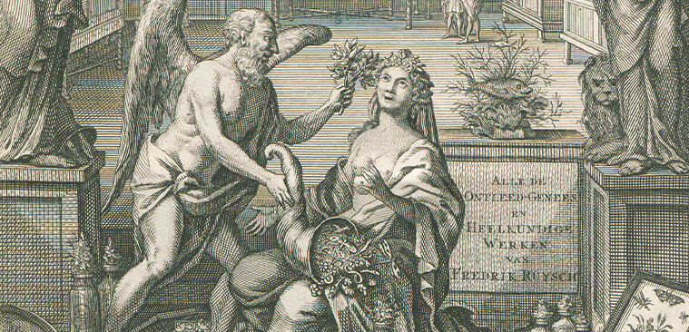
“Alle de ontleed- genees- en heelkindige werken …,” Frederik Ruysch, 1744
close next previous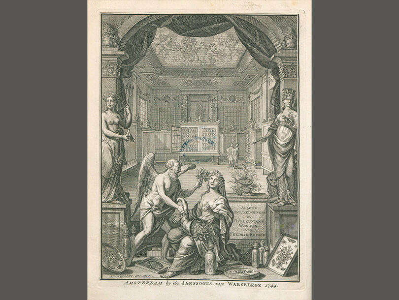
Dutch botanist and anatomist Frederik Ruysch (1638–1731) became known for his knowledge of anatomic preservation techniques and for his dioramas that incorporated human and earthly objects. Alle de ontleed- genees- en heelkindige werken is the Dutch translation of his Opera omnia anatomico- medico- chirurgica (1737), which was originally published in 1721. The three volumes highlight the human body, particularly organs and the anatomy of human babies and fetuses. The final section features animal and plant anatomy. His captivating and creative illustrations contain artistic, gruesome, and whimsical elements.

Historia de la composicion del cuerpo humano, Juan Valverde de Amusco, 1556
close next previous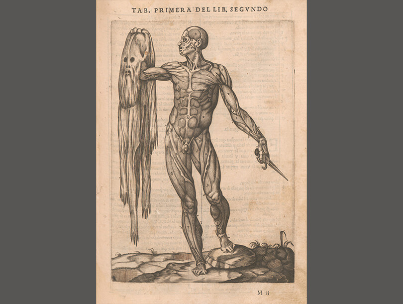
One of the first anatomy books published in Rome in the Spanish language, this became Juan Valverde de Amusco’s (ca. 1525–ca. 1588) most famous work. Like most anatomists during the period, Valverde compared existing works, performed his own dissections, and created new texts based on combining old and new information. The plate showing the flayed cadaver holding its own skin is one of Valverde’s most well-known images.
Anatomia del corpo humano, Juan Valverde de Amusco, 1559
close next previous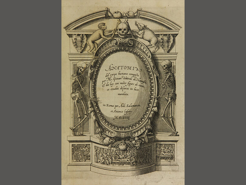
In this Italian translation of Juan Valverde de Amusco’s (ca. 1525–ca. 1588) Historia de la composicion del cuerpo humano (1556), the chapters remain the same, but the order of the plates is slightly different. The 350-page book is a compilation of seven chapters that discuss the main elements of the human body. The majority of the copperplate illustrations were taken from Andreas Vesalius’s (1514–1564) De humani corporis fabrica libri septem (1543).
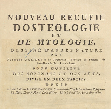
Nouveau recueil d'ostéologie et de myologie: dessiné d'après nature, Jacques Gamelin, 1779
close next previous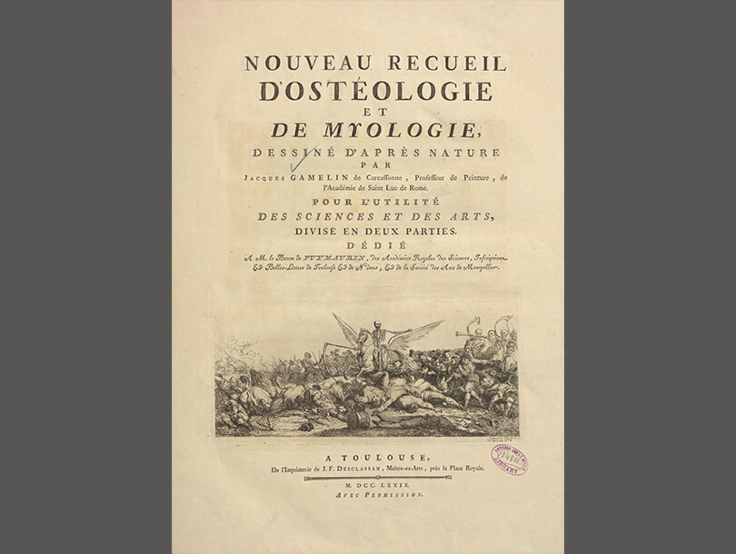
Jacques Gamelin (1738–1803) became a successful artist through the encouragement of his employer, a French industrialist, who recognized that Gamelin’s artistic promise far exceeded his business acumen and sent him to an art academy. Gamelin painted and engraved popular battle scenes, but his best-known work is Nouveau recueil d'ostéologie et de myologie, a two-part collection that includes anatomical scenes with mischievous skeletons at play, in religious and war vignettes, and many with Death at the helm. Included in this work is an engraved portrait of his lifelong patron, Baron de Puymaurin, to whom he dedicated the book. Gamelin also served as a painter to Pope Clement XIV.
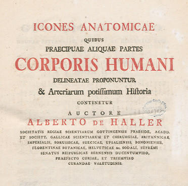
Icones anatomicae quibus praecipuae aliquae partes corporis humani delineatae proponuntur & arteriarum potissimum historia continetur ..., Albrecht von Haller, (anatomist); Rollinus, C. J. (artist), 1756
close next previous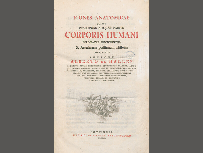
The successors of Swiss anatomist, botanist, and physiologist Albrecht von Haller (1708–1777) considered him to be the founder of a new epoch in anatomy and physiology. The eight parts published in 3 volumes reflect Haller’s precise investigations, corrections, and attention to detail in the entire arterial system of the body. This work served as an essential source about arteries and organs for more than a century.

Bernardi Siegfried Albini Tabulae sceleti et musculorum corporis humani, Bernhard Siegfried Albinus, 1749
close next previous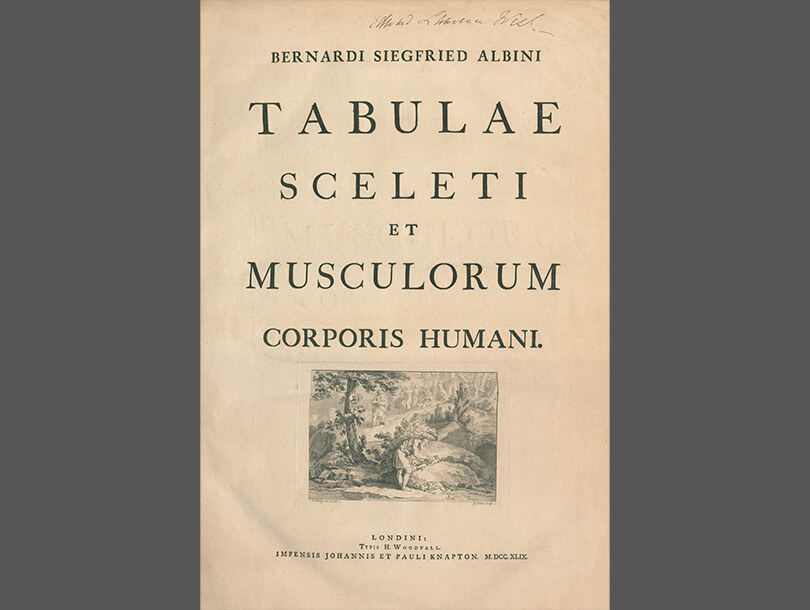
Famous for his studies of bones and muscles, surgeon and teacher Bernard Albinus (1697–1770) produced most of his work with Dutch illustrator and engraver Jan Wandelaar (ca. 1690–1759). In Tabulae, the two devised a new technique for rendering precise illustrations by placing nets with square webbing at intervals between the artist and the specimen in order to create a grid pattern for increased accuracy. Artistry also prevailed as many of the anatomical illustrations are in pastoral settings decorated with cherubim, wild animals, tropical plants, and vast landscapes.

Medical anatomy; or, Illustrations of the relative position and movements of the internal organs, Francis Sibson, 1869
close next previous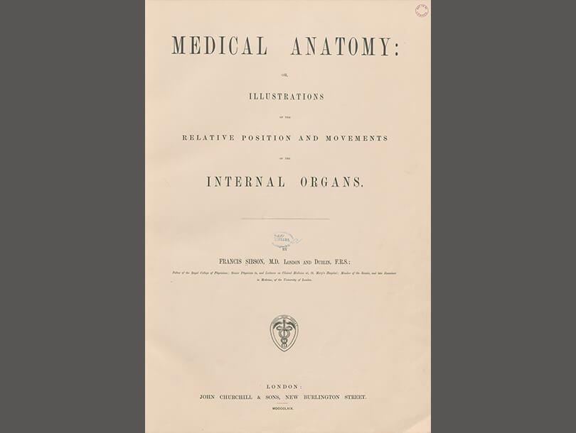
English physician and anatomist Francis Sibson (1814–1876) focused his anatomical studies on organs, particularly the heart and lungs. Medical Anatomy stressed the difference between healthy and diseased bodies, and how organs move and exist in living bodies—in contrast to the cadavers students typically examined. Sibson emphasized the differences that can be found between one body and the next and explained the natural and pathological reasons for such anomalies and variety among specimens. This volume contains 21 color plates and more than two dozen smaller drawings.
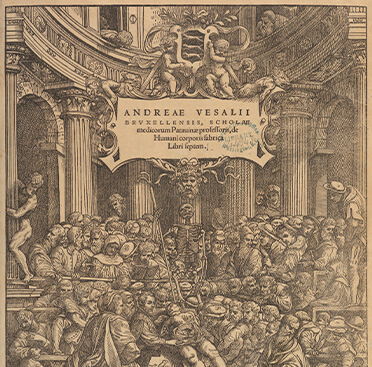
[Andreae Vesalii Bruxellensis, scholae medicorum Patauinae professoris] De humani corporis fabrica libri septem …, Andreas Vesalius, 1543
close next previous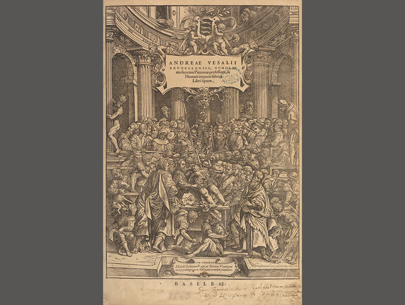
Published when he was 28 years old, this work brought Brussels-born anatomist Andreas Vesalius (1514–1564) immediate fame and prominence, as soon after, he was appointed physician to the Holy Roman Emperor Charles V. Accompanying the text are more than 250 anatomical illustrations printed from detailed woodcuts far superior to the drawings in other physiology atlases of the time. Among the illustrations are the frontispiece of Vesalius himself at the center of a crowded anatomy theatre.

Anatomie du gladiateur combattant, applicable aux beaux arts; ou, Traité des os, des muscles, du mécanisme de mouvemens, des proportions et des caractères du corps humain, Jean Galbert Salvage, 1812
close next previous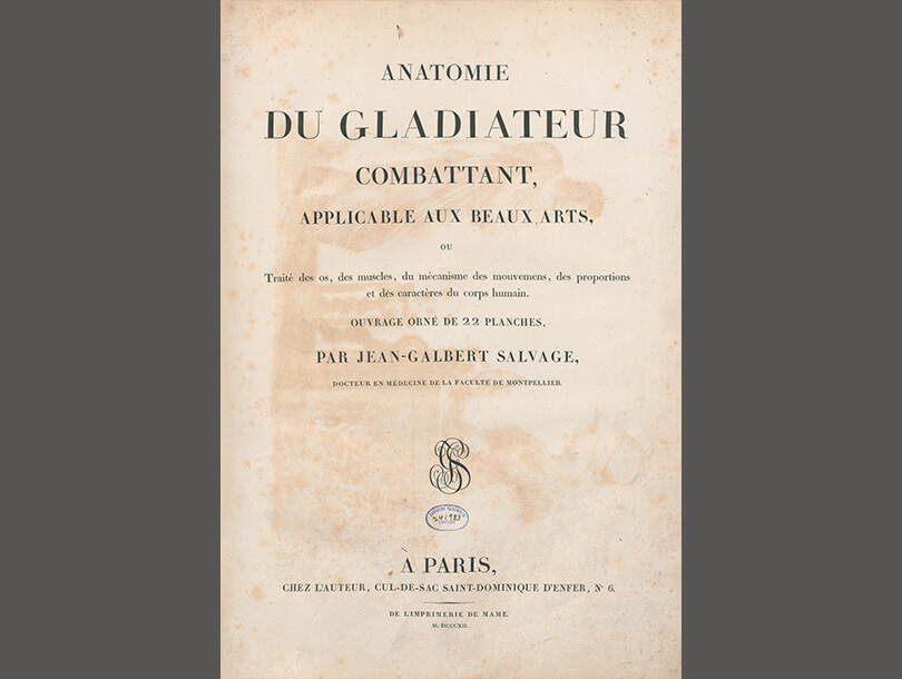
A military doctor in Napoleon’s army, physician Jean Galbert Salvage (1770–1813) earned a post in Paris after excellent service. Once there, Salvage developed his artistic skills to create Anatomie du gladiateur combattant as the subject of his anatomy text. By using the Borghese Gladiator, an ancient Roman statue that had recently been purchased by Napoleon and on display in the Musée Napoleon, Salvage blended the best anatomical knowledge with what was considered the most revered art and sculpture in the Western world at that time. The drawings show the Gladiator from all angles with his muscles and bones anatomically rendered. These particular anatomical studies represent a genre within fine art as well as being instructive.
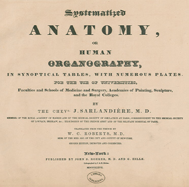
Systematized anatomy; or, Human organography (second edition), Jean Baptiste Sarlandière, 1837
close next previous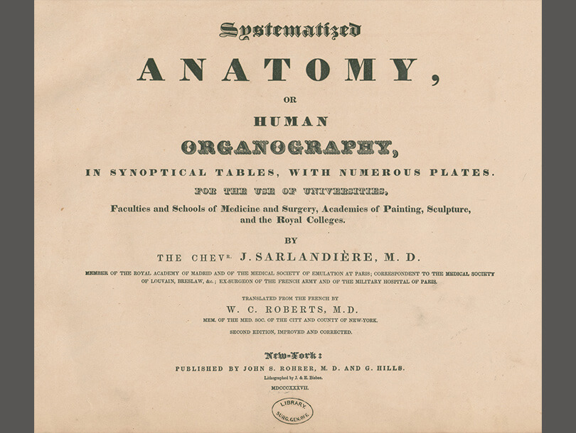
In 1837, American doctor W. C. Roberts (1810–1873) translated and printed 1831 Anatomia methodica by Jean-Baptiste Sarlandière (1787–1838). Sarlandière hoped that this small, reference-like volume might be a useful guide for physicians, students, and artists alike. The 15 leaves of color lithographed plates each depict collections of human physiology: muscles (myography), bones (osteography), organs (splanchnography), nerves (neurography) and the circulatory system (angiography). Both the color and the meticulous identification notations make the publication an inviting and captivating treasure.

Tabulae anatomicae ligamentorum corporus humani, Florian Caldani, 1803
close next previous
Floriano Caldani (1772–1836), physiologist, anatomist and naturalist, was the nephew of physician Leopoldo Caldani and would succeed his uncle as the chair of anatomy at the University of Padua in Italy. The two men collaborated to create one of the most famous anatomical books of the time, Icones anatomicae, which was used widely in Europe for decades. This smaller volume focuses on human ligaments. Though the work is not considered groundbreaking, the illustrations show Floriano Caldani’s devotion to the descriptive canons of classical anatomy. He dedicated the volume to his uncle, Leopoldo Caldani.

Topographisch-anatomischer Atlas. Nach Durchschnitten an gefrorenen Cadavern, Wilhelm Braune, 1872
close next previous
Unique to anatomical studies at the time, this atlas by Christian Wilhelm Braune (1831–1892) contains illustrations drawn from a frozen human cadaver that had been sliced with a very fine-toothed saw. This technique provided a view of the body’s interior from a horizontal perspective. Braune hoped that his illustrations would increase surgical knowledge, especially the location of organs in relation to each other. In addition, Braune studied the physiology of the human gait—the biomechanics of gravity and balance. The illustrations he created from these studies helped establish an understanding of the center of gravity in the body. The volume begins with Braune’s acknowledgement of the young man whose body he dissected—a physically healthy 21-year-old who had hanged himself—and the process by which he froze the young man’s body for dissection.

Elementi di anatomia fisiologica applicata alle belle arti figurative (Atlas), Francesco Bertinatti, 1839
close next previous
Bertinatti’s publication consists of two volumes of text and an atlas with illustrations of anatomical studies. The realistic anatomical drawings, at times, are set in an imaginary setting.
Bartholomaei Eustachii anatomici summi Romanae archetypae tabulae anatomicae novis explicationibus, Bartolomeo Eustachi, 1783
close next previous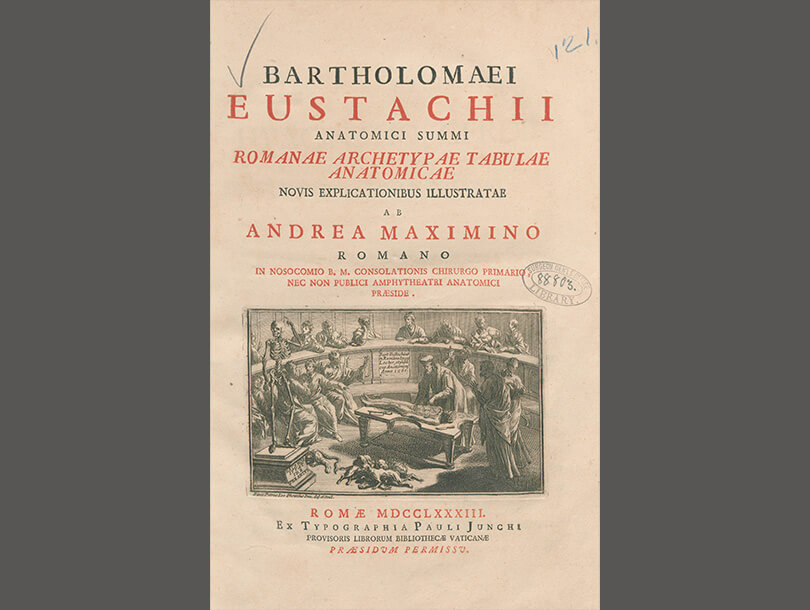
This 1783 edition includes an etching of the author, Bartholomeo Eustachi (d. 1574) teaching and dissecting in an anatomy theatre on the title page. The plates feature a unique identification system—the use of numbered rulers on the edges of each plate to allow for easy in-text identification without adding numbers or letters next to the image. Born sometime at the beginning of the 16th century, Bartolomeo Eustachi is among the early founders of anatomical studies, and he contributed essential knowledge to human physiology. He significantly increased knowledge about the inner ear (thus the Eustachian tube bears his name), discovered the adrenal glands, and wrote the first in-depth study of teeth. He served as the physician to the Duke of Urbino and later to the Cardinal Giulio Della Rovere in Rome. Although completed in 1552, due to the religious censoring of anatomists’ work during the Renaissance, this volume was hidden in the papal library until its first publication in 1714.
Anatomie des parties de la génération de l'homme et de la femme: représentées avec leurs couleurs naturelles, selon le nouvel art, jointe a l'angéologie de tout le corps humain, et a ce qui concerne la grossesse et les accouchemens, Gautier d'Agoty, 1773
close next previous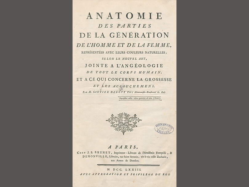
This thin volume is remarkable in its use of a color printing method based in mezzotint and etching techniques. The eight color plates in this volume may be combined in pairs to create four larger images. All but one feature the anatomy of pregnant and birthing women. French artist Gautier d’Agoty (1717–1785) studied for many years with the painter and engraver Jacob Christoph Le Blon. The two men later quarreled over which one of them had invented the color printing method both used. Gautier d’Agoty’s prints convey a distinct painterly quality, and the specimens are often posed in the typical 18th-century portrait style, calmly gazing back at the viewer despite their lack of skin, exposed muscles, and organs.
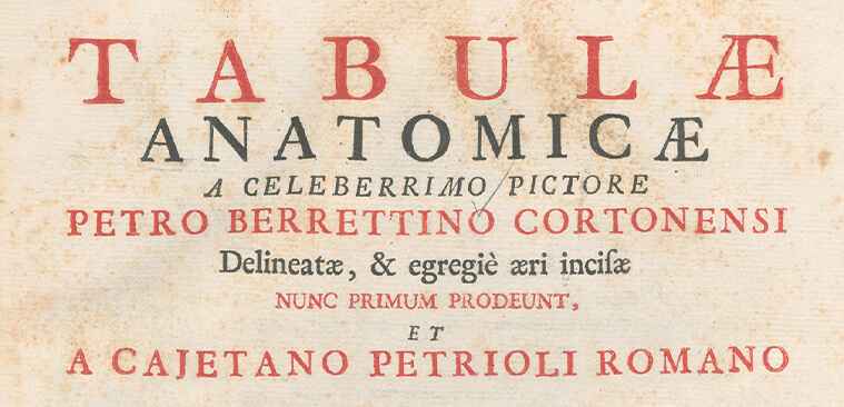
Tabulae anatomicae ... delineatae, & egregie aeri incisae nunc primum prodeunt, et a Cajetano Petrioli ... notis illustratae., [Berrettini] da Cortona Pietro, 1741
close next previous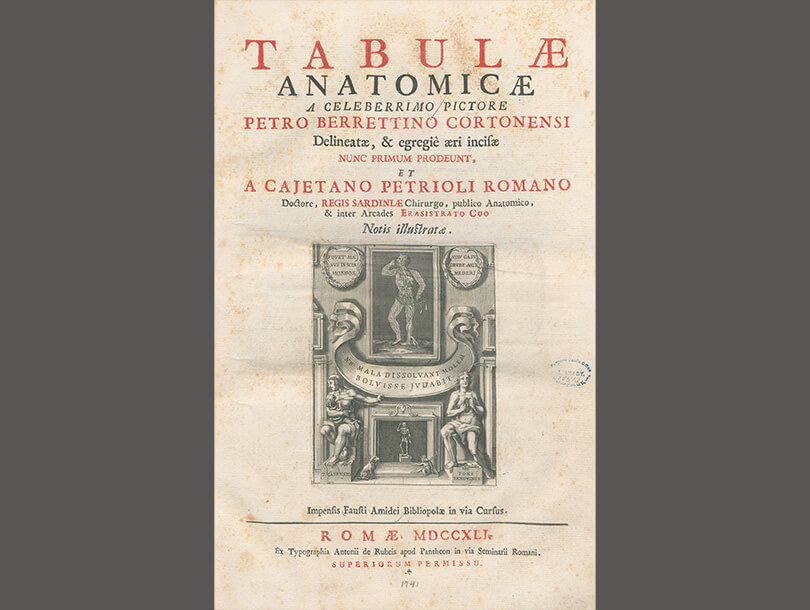
Italian artist Pietro da Cortona (1597–1669) produced the original illustrations for this anatomical atlas in the 17th century, and Gaetano Petrioli modified the illustrations for publication some 70 years after Cortona’s death. The 19 male figures are all posed in a classical Greek and Roman fashion, often with columns and other architectural details of the Classical Antiquities era popular among artists of Cortona’s time. The illustrations focus primarily on muscles, nerves, and blood vessels. Several of the dissected figures shown in the illustrations hold mirrors or other oval shapes within which are details of related aspects of the larger anatomical drawings. Petrioli modified each plate by embellishing the scenes with smaller illustrations to fill the entire space available.

Anatomia universale del Paolo Mascagni, Paolo Mascagni, 1833
close next previous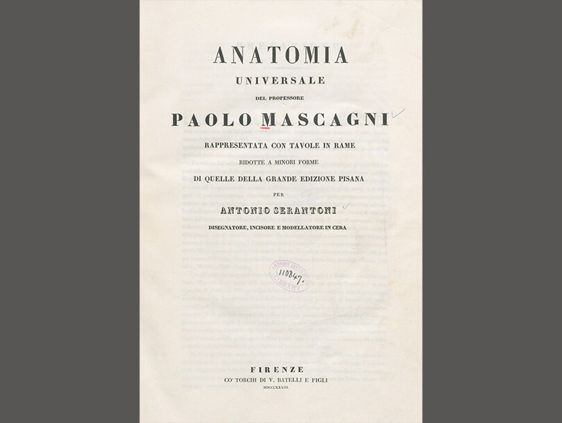
Italian physician Paolo Mascagni (ca. 1752–1815) is best known for providing the first systematic and complete description of the lymphatic system in 1787. The publication of Vasoum lymphaticorum brought him fame in Europe and led to a professorship in Pisa and membership in two medical academies. Fifteen years after his death three professors at Pisa compiled and printed Anatomia universale. This 600-page atlas is a comprehensive study of anatomy that includes 44 hand-colored plates. The careful shading and rendering of the etching, along with the use of color, makes some of the images appear three-dimensional.
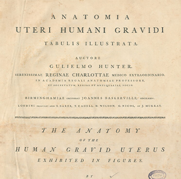
Anatomia uteri humani gravidi tabulis illustrata/ The anatomy of the human gravid uterus exhibited in figures, William Hunter, 1774
close next previous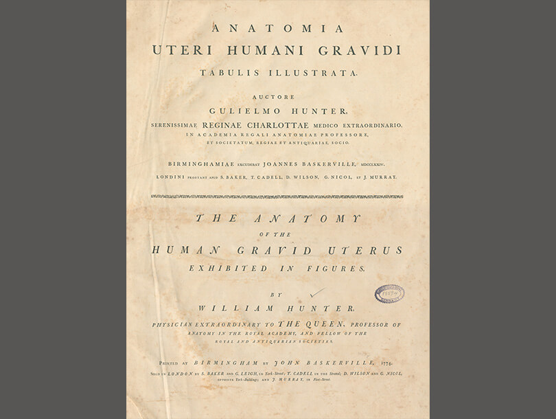
Scottish anatomist and physician William Hunter (1718–1783) spent much of his career studying pregnancy, labor, and the gravid uterus. Although his younger brother John was more well-known as a physician, William’s work on the anatomy of the pregnant uterus brought attention to a subject largely ignored prior to his studies. In 1751, he seized upon an opportunity—the pregnant woman whose body is depicted died suddenly and at a time of year when cold weather allowed for dissections. This work broke new ground in its description of the musculature of the uterus and the uterine lining. Hunter also discovered that the fetal-placental circulation was completely independent of that of the mother. Written in both Latin and English, the succinctly written volume contains 34 exquisitely detailed illustrations by Dutch engraverJan van Rymsdyk (fl. 1750–1788)
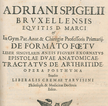
Adriani Spigelii Bruxellensis equitis D. Marci ... De formato foetu liber singularis : aeneis figuris exornatus : epistolae duae anatomicae : tractatus de arthritide : opera posthuman, Adriaan van de Spiegel, 1626
close next previous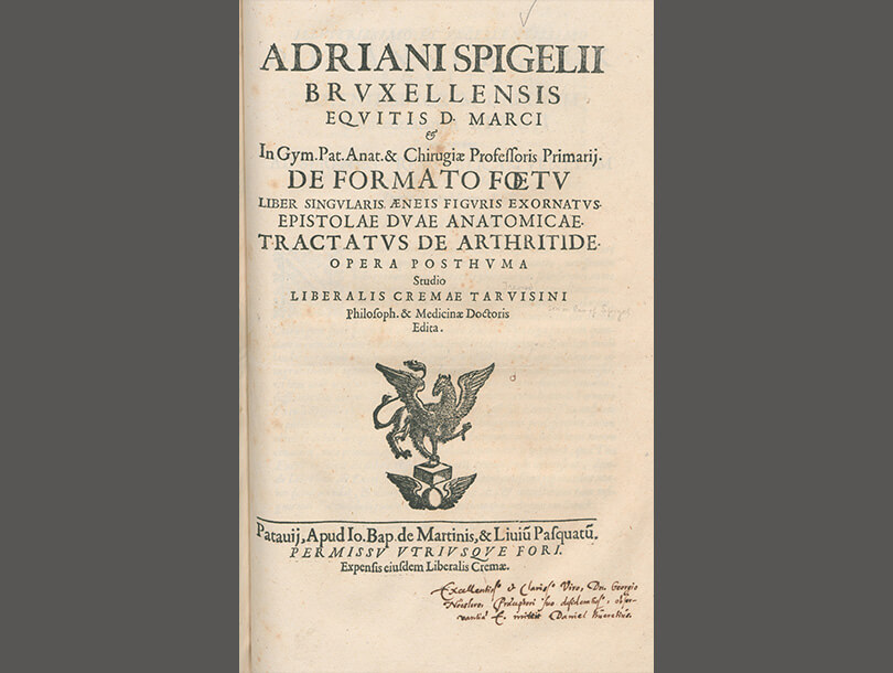
This volume ultimately brought together the work of two physician-anatomists, an artist, and an engraver. After Adriann van de Spiegel’s death, Liberalis Crema, Spiegel’s son-in-law, wanted to posthumously publish Spiegel’s unillustrated text, De formato foetu liber singularis. To accompany the text, he obtained nine copperplate engravings that anatomist Guilio Cessari had previously completed on the topic of fetal development. These illustrations are the work of Titian’s student, Odoardo Fialetti, and engraver Francesco Valesio. They show graceful pregnant women in various stages of uterine dissection with wombs, newborns, and placenta, all artistically conveyed.
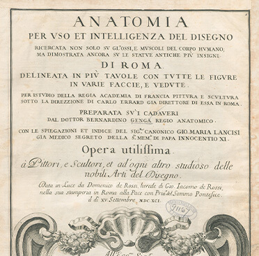
Anatomia per uso et intelligenza del disegno ricercata non solo su gl'ossi, e muscoli del corpo humano: ma dimostrata ancora su le statue antiche più insigni di Roma : delineata in più tavole con tutte le figure in varie faccie, e veduta, Bernardino Genga, 1691
close next previous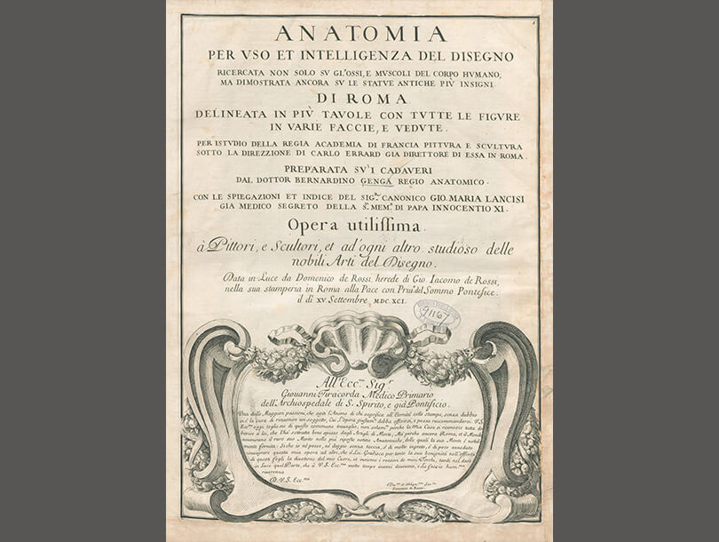
Published one year after he died, Italian scholar and artist, Bernardino Genga’s (1620–1690) Anatomia per uso et intelligenza del disegno features 59 copperplate engravings of bones and muscles, as well as drawings of famous antique sculptures from a variety of angles. A scholar of Classical medical texts—Hippocrates in particular—Genga was also adept in the preparation of anatomical specimens. He taught anatomy to artists at the French Academy and practiced surgery in the Hospital Santo Spirito in Sassia, both in Rome.

Ontleding des menschelyken lichaams, Govard Bidloo, 1690
close previous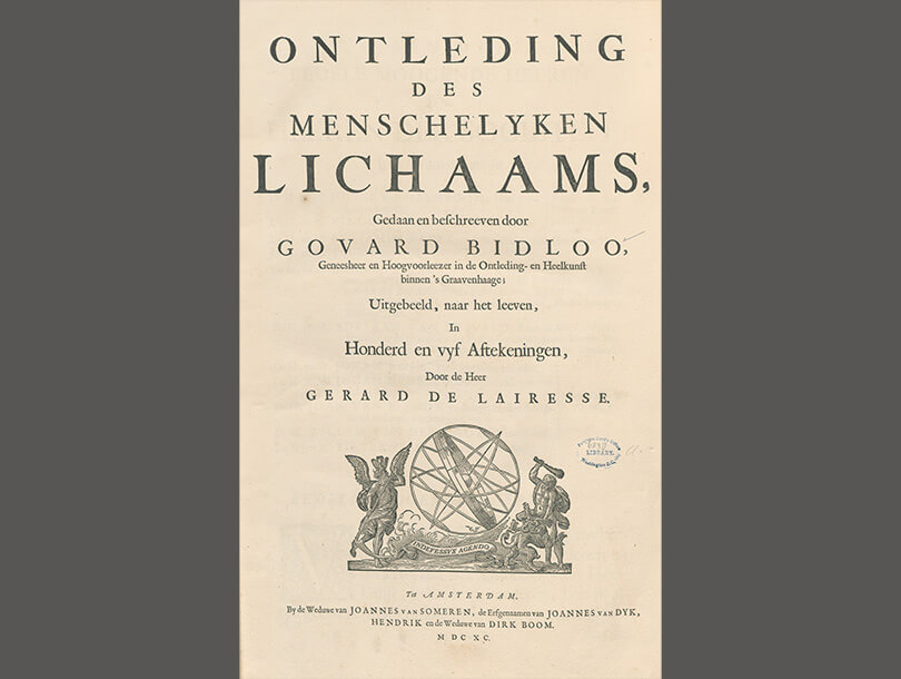
Govard Bidloo (1649–1713) was a multi-talented Dutch physician who studied surgery, lectured in anatomy, and was the head of the national hospital service in Amsterdam and then later in England. Ontleding des menschelyken lichaams, also known as Anatomia Hvmani Corporis, is generously illustrated with 105 plates by Dutch artist Gerard de Lairesse (1640–1711). Bidloo described the papillary ridges on fingers in this atlas, thus laying the foundation for later forensic identification techniques. English surgeon William Cowper reprinted an expanded English version of this atlas in 1698 but failed to give Bidloo or Lairesse proper credit. This began a well-known plagiarism dispute a decade before the legal concept of copyright was implemented.